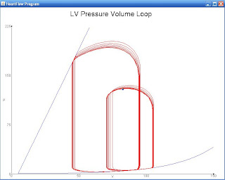
Let's just start with a quick look at what a PV Loop looks like:
There it is in its glory, Not much to look at, eh? Don't worry! With the use of my laptop, let's get a few things labelled so that the PV Loop is a lot less mystery and a lot more awesomely useful.
Let's start with the axes:
Just for clarity, the x-axis depicts volume, while the y-axis depicts pressure.
This line shown in the PV Loop is a depiction of contractility. Shortly, you will get a chance to see how a change in contractility (via that line) alters the shape of the PV Loop.
The width of the PV Loop corresponds to stroke volume. Just like contractility, you will shortly see how easily the stroke volume can be altered by the shape of the PV Loop.
Now, for big concepts: A PV Loop depicts the events of a single, hypothetical heartbeat. What is also true is that the PV Loop is not a 'snapshot' of the heart. To read the PV Loop, you must travel along the PV Loop. Does that make sense? What I mean is, you pick a point to start (let's say the Upper Left corner, and then follow the loop's line. Something that is true is that throughout that heartbeat, the pressure is always changing. As you follow the line of the PV Loop, you never just stay at the same value along the y-axis (the axis that depicts pressure). You may return or have similar pressure value eventually, but you are just stuck at one particular value of pressure. How does this apply to the x-axis (the axis that depicts volume)? Excellent! There are two points in time in which the volume in the heart is unchanging (depicted by the two vertical lines, one on the left and another on the right of the PV Loop). There are two points in time (within the heart) that the volume is unchanging, one (the left) value is isovolumetric relaxation and the other (the right value) is isovolumetric contraction. We will come back to both, shortly.
Here are some of the major events within the left ventricle. Remember, that the PV Loop can depict activity in any of the four chambers of the heart. In our example, we are talking specifically about the PV Loop in the Left Ventricle. Let's start in the Upper Left corner of the loop. At this moment, I have denoted that the aortic valve closes. The aortic valve is the valve that exists in the aorta, and can allow or prevent communication between the LV and the body. As a result of the aortic valve closing, the chamber exists in a state in which no valves are open. I will follow with a labelled picture, but the left vertical line of the PV Loop depicts, then, a time of isovolumetric relaxation. Picking apart the word, isovolumetric relaxation refers to a period of relaxation in the heart in which there is no change of volume occurring (a result of the fact that no heart valves are open).
At the Bottom Left corner of the PV Loop, the mitral valve opens. Remember, the mitral valve is the valves that separates the left atrium from the left ventricle. What do you suppose happens when the mitral valve opens and allows communication between the LA and the LV? Of course! When the mitral valve opens blood is allowed into the left ventricle and diastolic filling of the chamber begins! Great job!
At the Bottom Right corner of the PV Loop, the mitral valve closes. Here we are again, with none of the heart valves that communicate with the LV being open. Again, no change in volume occurs, making this isovolumetric. This time, however, rather than relaxation occurring, contraction is occurring. What's the difference, or what makes one relaxation and the other contraction? I'm glad you asked! So, let's start with isovolumetric contraction (the right vertical line). What has occurred in the heart (due to diastolic filling)? It's filled with blood! Something we have yet to discuss is that there is electrical activity going on within the heart (preparing the heart to contract for a heartbeat). So, as the cardiac myocytes begin to contract (or, shrink in size), that pressure, combined with the pressure created by the stretching of cardiac myocytes due to being filled with blood, continually increases. In contrast, isovolumetric relaxation (the left vertical line) is occurring after contraction (or ejection of blood from the heart) has occurred. As you can imagine, that is a huge relief or a relaxing event for the heart. It's allowed to return to its normal, original size. I will be providing a labelled diagram of this in just a sec, but I think the next paragraph will help here.
Finally, at the Upper Right corner of the PV Loop, the aortic valve opens. Now, what happens in the heart? Right! When the aortic valve opens (alongside that contraction of the left ventricle) blood is ejected from the heart to the aorta (to be carried to the body). Starting the Bottom Right corner and ending with the Upper Left corner, we have described contraction (or the period of systole in the heart).
Now, for some labelled diagrams:
And, to just be even more helpful (I know my artwork is terrible), here's a professional depiction of the PV Loop, with all of the bits and pieces labelled (I just wanted to go slow, initially). If you are confused (as I feel that I may not have done as well as I could have, please leave a comment):
Here's a fairly isolated change in Preload:
What changes do you see between the normal loop (more to the left) and the loop with increased preload (more to the right)?
Did you see any of these changes? As noted, an increase in preload is going to result in an increase in stroke volume! But, we already knew this, and we certainly didn't need a PV Loop to explain the relationship. If preload were to increase, the amount of blood that fills the left ventricle during diastole is going to increase (see the notation of the end volume in appropriate colors). As a result, when that blood is ejected from the heart, more blood will be ejected (or a higher stroke volume will be ejected). What are some things that might increase preload? Well, fluid retention or fluid administration is an easy way to increase preload in the body. A decrease in preload (not depicted) might result from blood loss or even the use of diuretics (Lasix).
How about this fairly isolated change in afterload? What changes do you see in this PV Loop?
Did you see these changes:
So, what do we see with this fairly isolated increase in afterload. Again, stroke volume is affected. However, this time stroke volume has decreased in response to the increased afterload. But, then again, we already knew that too! If afterload were to increase, the force that the left ventricle would have to overcome to eject blood would also increase. Because of this, the LV is going to have to do more work in order to compensate for this increase in resistance. Note, again, the changes along the axis. On the y-axis, one can see that the initial SV results in smaller amount of pressure being exerted, when compared to the second PV-Loop. Certainly, that increased pressure reflects that increased resistance or load that must be overcome for blood ejection. What might cause an increase in afterload? Well, if we had taken this PV Loop measurement right as or fairly soon after the aorta had stenosed (or collapsed), we might see an increased afterload like this.
And our final loop, a change in contraction (thus far we've had no change in contraction):
Wow! There are some drastic changes in the loop with this change in contractility. What things do you see?
Some notes on the effects the change in contractility have had on this loop. First, the slope of the contractility line has decreased. The decrease in slope corresponds to a decreased contractility. Look at the stroke volumes. In the initial heart PV Loop, the SV is much greater than the stroke volume created by the second PV Loop. So, what might cause changes in contraction (such as these)? Well, let's say on the initial loop (the one to the left), it was taken while I was running from a monster (high contraction going on). The second loop (or the loop to the right) might have been taken after I had been given a sedative by a doctor, following my intense run in with a monster. Here, the sedative has decreased contraction in my heart.
There you have it! That was a whole lot about the PV Loop. If you are interested in a great simulator for the study of the heart, here is a FANTASTIC website: http://ccnmtl.columbia.edu/projects/heart/sim.html. Here you will find the method that allowed me to make this post.
The PV Loop, once you understand and get past the WOW factor (and by WOW, I mean, "WOW, what is this mumbo jumbo??"), can be quite helpful in making diagnoses. Something that I have yet to note (or I should say stress) is that the PV Loop makes no consideration for an increase in wall thickness. It couldn't, right? The PV Loop is a measurement of only one heartbeat. As we have seen, wall thickness is something that changes over a long period of time. Also in need of note, while I proposed to you some 'isolated' changes in some of the values, this really is not going to happen in the heart. Why? Well, that's because the heart has those Top 3 Priorities, and it is always changing things (all things) to ensure that those priorities are met.
Cheers (and believe me when I say, this will definitely need a reread once I have slept some)!
** Reread and improved at 5:00 PM the next day **


No comments:
Post a Comment